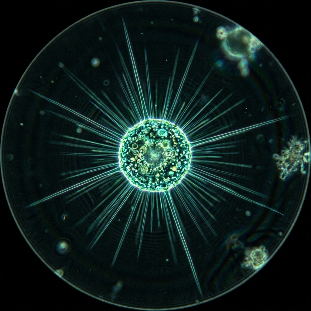
 Figure 1: Actinophrys floating with long, thin axopodia extending in all directions like a sun (*Image is a conceptual CG render)
[!NOTE] *All microorganism images used in this article are conceptual 3D CG renders.
🎯 Quick Summary & FAQ (Conclusion First)
Before diving into the detailed guide, let’s quickly answer some of the most common questions about Actinophrys (Sun Animalcules).
Q. What kind of organism is Actinophrys (Sun Animalcules)? A. It is a single-celled organism, nearly invisible to the naked eye, with countless needle-like spines (axopodia) radiating from its body. Because its appearance resembles the sun’s corona or rays, it is commonly called “Sun Animalcules” (Heliozoa). A close relative of amoebas, it is a microscopic hunter that uses these needle-like pseudopodia to snag passing plankton.
Q. Can I culture (breed) them at home? A. Yes, it is possible. However, you cannot culture them using only Ebios tablets like Paramecium. Since Actinophrys is a pure predator that only feeds on live prey (other single-celled organisms), you must culture small flagellates or ciliates (such as Paramecium or dinoflagellates) either simultaneously or in advance to serve as food.
🔬 1. The Amazing Ecology of Actinophrys: Beautiful Axopodia and Fusion Phenomena
Actinophrys (primarily the freshwater species Actinophrys sol) possesses one of the most elegant geometric forms in the plankton world.
How the Needle-like Weapon “Axopodia” Works
The countless needles extending from the Actinophrys are not merely decorative; they are cytoplasmic projections called axopodia. Inside them, thin protein fibers called “microtubules” are tightly bundled together, giving the needles their rigid structure. The surface of the axopodia is covered with an adhesive substance and contains toxins (extracellular toxins) that paralyze prey on contact. Once prey touches these needles, it is immobilized. Actinophrys then retracts its axopodia or extends surrounding cytoplasm to envelop the prey, slowly drawing it inside the cell body to form a food vacuole.
The Astonishing “Cell Fusion” Phenomenon
One of the most unique ecological traits of Actinophrys is cell fusion when capturing large prey. When encountering a ciliate much larger than itself, or when multiple prey are caught at the same time, nearby Actinophrys individuals will temporarily fuse together to form a single, giant multinucleated cell. After jointly enveloping and digesting the prey, they divide back into their original sizes and separate once digestion is complete. This dynamic cooperative hunting (if it can be called that) is one of the most exciting phenomena to observe under a microscope.
| Feature | Actinophrys (Sun Animalcule) | Amoeba (Common Amoeba) |
|---|---|---|
| Pseudopodia Shape | Rigid, needle-like “axopodia” | Fluid, irregular “lobopodia” |
| Movement | Mostly floats suspended or rolls slowly | Crawls on surfaces by extending pseudopodia (amoeboid movement) |
| Feeding Style | Ambush (captures prey that touches needles) | Active pursuit/engulfing (surrounds and phagocytoses) |
| Cell Fusion | Fuses temporarily during feeding or harsh conditions | Generally does not fuse |
🧪 2. How to Collect and Culture Actinophrys at Home
Here are the practical techniques to find Actinophrys in nature and maintain them at home.
📦 Materials Needed
- Collected pond water and sediment (near decomposing leaves is best)
- Prey plankton (Paramecium or small flagellate culture)
- Dechlorinated tap water or commercial soft mineral water
- Small plastic petri dishes or clear lidded containers
- Fine-tip pipette (dropper)
- Sponge or filter paper (for pipette filtration)
Collection Approach from Streams and Ponds
Actinophrys prefers slow-moving ponds, swamps, marshes, or around aquatic plants and fallen leaves in rice paddies.
- Select Target Area: Target shallow waters with abundant aquatic vegetation and accumulated organic sediment.
- Sampling: Collect some bottom mud, decaying leaves, and aquatic plants along with water in a plastic bottle or jar.
- Settling: Open the lid of the container and let it sit on a bright windowsill (avoiding direct sunlight) for about half a day. Actinophrys will gather around the aquatic plants near the water surface or the walls of the container, drawn by light-seeking prey or due to their own gentle phototaxis.
🌿 The “Two-Step” Culturing Method for Actinophrys
Like amoebas, Actinophrys does not feed directly on bacteria; instead, it feeds on the “small plankton” that eat bacteria. Therefore, you establish the following food chain:
graph LR
    A[Ebios Tablet / Rice Grain] --> B(Bacteria)
    B --> C(Paramecium / Small Flagellates)
    C --> D((Actinophrys))
Step 1: Secure the Food Supply (Paramecium, etc.)
Pre-culture Paramecium or Chilomonas (small ciliates/flagellates) in advance. Dissolve 1/4 of an Ebios tablet in 500 ml of dechlorinated water, add a starter culture, and let it sit for a few days until the liquid becomes cloudy (indicating a high density of bacteria and Paramecium).
Step 2: Isolating and Inoculating Actinophrys
- Locate Actinophrys in the collected water under a microscope.
- Use a fine-tip pipette to isolate and transfer several to a dozen individuals into a petri dish containing clean water (this process is called “picking” or “loading”).
- Add a very small amount (a few drops to about 1 ml) of the food culture prepared in Step 1.
Step 3: Maintenance and Subculturing
Store in a cool place (15°C - 22°C) away from direct sunlight. Too much food will cause the water to spoil and kill the Actinophrys. Check under the microscope every few days and add a few drops of the prey culture as the food supply decreases. About once every two weeks, fill a new petri dish with water and transfer a few individuals with a pipette to subculture them into a fresh environment.
🔬 3. Microscopy Hacks for Beautiful Observations
Since the axopodia of Actinophrys are extremely thin, they can be transparent and difficult to see under standard brightfield observation. Use the following hacks to maximize visible details.
1. Darkfield-Style Lighting DIY
To make the Actinophrys needles glow beautifully, darkfield microscopy—which illuminates the specimen from an angle—is ideal.
- DIY Darkfield Filter: Place a transparent plastic sheet with a black circle (about 1 cm - 1.5 cm in diameter) drawn in the center under the microscope’s condenser (above the light source) to act as a patch stop.
- Effect: The background turns completely black, and the round cell body of the Actinophrys, along with its countless radiating axopodia, glows white like a star floating in space.
2. The “Spacer” Hack to Prevent Crushing the Axopodia
Actinophrys is a three-dimensional sphere. Placing a cover slip directly onto the slide will crush the cell under its weight, breaking all the axopodia.
- Hack Method: Create a small gap (spacer) by sticking thin strips of cellophane tape or tiny pieces of plastic film on the corners (or left and right edges) of the slide before placing the cover slip.
- Result: This maintains an adequate depth in the water layer, preventing the Actinophrys from being crushed and allowing you to observe them floating and swimming while fully expanding their axopodia in 360 degrees.
3. Capturing the Moment of Predation
After placing the Actinophrys on the slide, add a drop of concentrated Paramecium culture to the edge of the cover slip, and draw water out from the opposite side using a piece of paper towel to guide the Paramecium toward the Actinophrys. The moment a Paramecium touches the Actinophrys needles, it stops dead in its tracks. You can then watch the thrilling, real-time hunting scene as the axopodia slowly retract and pull the prey in.