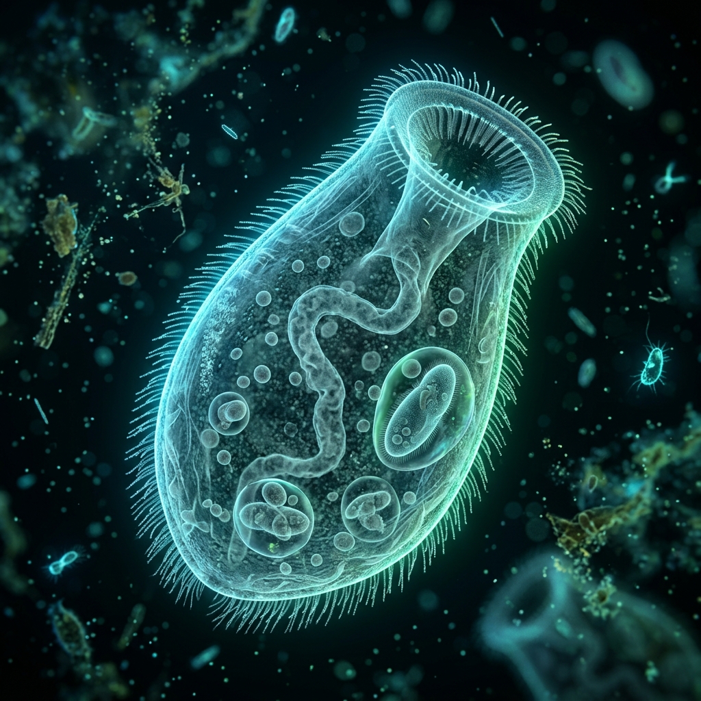
 Fig 1: A Bursaria with a massive, pouch-like oral groove (vestibulum), swallowing a paramecium (*Conceptual image).
[!NOTE] *All microorganism images used in this article are conceptual 3D CG renders.
🎯 Quick Summary & FAQ (Key Takeaways)
Before diving into the detailed guide, here are quick answers to the most common questions about collecting and culturing Bursaria.
Q. Can you see Bursaria with the naked eye?
A. Yes, absolutely!
Since they grow to 0.5 mm–1.0 mm (nearly 1000 μm) in length, they are easily visible to the naked eye as tiny white specks swimming and slowly rotating in the water, despite being single-celled organisms.
Q. What is the difference between Didinium and Bursaria?
A. They differ in size, diet, and hunting style.
Didinium feeds exclusively on Paramecium, paralyzing it with toxicysts before swallowing. Bursaria, on the other hand, is several times larger and sucks in not only Paramecium but also rotifers and other protozoans like a vacuum cleaner using its large, pouch-like vestibulum (oral groove) to swallow them whole.
🔬 1. The Amazing Biology of Bursaria
Known among microscopy enthusiasts as the “whale of the micro-world,” Bursaria (scientific name: Bursaria truncatella) is a giant ciliate. While it belongs to the same ciliate group as Paramecium, its scale and predation style are on a completely different level.
A Giant Vestibulum That Vacuums Up Prey
The most defining feature of Bursaria is the massive, slit-like cavity (vestibulum) occupying almost half of its body. The inside of this pocket is lined with dense, specialized cilia that generate a powerful water current, vacuuming in surrounding water along with prey into its cytostome (cell mouth).
Identification Key: The Bead-like Macronucleus
When observing Bursaria under a microscope, you can see a long, ribbon-like (or bead-like) macronucleus extending in a “C” or “S” shape inside the body. This distinctive nuclear structure, combined with its pouch-like silhouette that looks almost cut in half, makes it easy to distinguish from other large ciliates like Stentor.
Microorganism Size & Feature Comparison
| Organism | Average Size | Key Morphological Features | Primary Diet | Predation Style |
|---|---|---|---|---|
| Bursaria (This species) | 0.5 mm - 1.0 mm | Giant pouch/purse shape, translucent with a bead-like macronucleus | Paramecium, rotifers, large single-celled organisms | Sucking in & swallowing whole via water currents |
| Paramecium | 0.15 mm - 0.25 mm | Slipper-shaped, elongated and streamlined, swims rapidly | Bacteria, microalgae | Filter feeding via oral groove cilia |
| Stentor | 0.5 mm - 2.0 mm | Trumpet-shaped (highly contractile), blue-green or colorless, sessile ability | Organic detritus, single-celled algae | Drawing in prey via ciliary ring currents |
| Didinium | 0.1 mm - 0.15 mm | Barrel-shaped, two ciliary bands around the body with a snout | Paramecium only (specialist) | Paralyzing prey with toxicysts (harpoons) and swallowing whole |
2. Collection Methods & Identification in Natural Waters
Bursaria can be found in ponds, marshes, wetlands, and rice paddies. They are highly adaptable. Your best bet is to target spots rich in organic matter where prey like Paramecium and rotifers thrive.
Collection Steps
- Target Locations: Scoop up water along with bottom debris/sediment from shallow pond edges, especially near aquatic plants or areas where fallen leaves accumulate and decay.
- Container Choice: Prepare a wide-mouth plastic bottle (approx. 1 liter). Fill it about halfway with sediment and water, leaving the other half as air space for transport.
- Settling & Scouting: Once home, let the bottle sit for a few hours until the mud settles. Direct the container toward room light and look through the side. Scan for “tiny white specks swimming slowly in the mid-water or right above the mud, spinning on their own axes.”
- Isolation: When you spot a white speck, use a fine-tip dropper to suck it up directly along with a bit of water. Transfer it to a petri dish filled with clean water and inspect it under a microscope.
3. Culturing Bursaria at Home (The Paramecium Feeding Method)
Because Bursaria are voracious eaters, keeping them in raw pond water without food will lead to starvation and complete die-offs within a few days. For a stable culture, you must set up a parallel breeding line of Paramecium to serve as prey.
Culture Setup & Daily Maintenance
- Preparing Base Water: Use aged, dechlorinated tap water or commercial bottled soft mineral water.
- Culture Vessels: Shallow containers with a large surface area, such as glass petri dishes or small plastic tubs, work best. Keep the water shallow (approx. 1.5 cm to 2.0 cm) to ensure optimal oxygen exchange.
- Feeding Frequency: Every 3 to 4 days, add a small amount of concentrated, home-cultured Paramecium (just enough to satisfy the Bursaria).
- Water Changes & Debris Removal: Once a week, use a pipette to clean out waste and debris from the bottom of the container. Replenish the lost water with fresh base water.
[!WARNING] Beware of Water Quality Deterioration from Overfeeding! If too much organic residue (such as yeast or rice bran from the Paramecium culture medium) enters the Bursaria container, anaerobic bacteria will bloom. This causes oxygen depletion, which can melt and kill the Bursaria. When feeding, try to concentrate and rinse the Paramecium with clean water before introducing them.
4. Microscope Hacks: Observing and Capturing the Dynamic Swallowing
Due to their immense size, the entire body of a Bursaria fits within the frame at a microscope’s lowest magnification (40x to 100x). This makes them an excellent target for smartphone photography and videography.
Slowing Things Down: The Glycerin/Methylcellulose Hack
While Bursaria are not as fast as Paramecium, they constantly rotate while swimming, making it easy to lose focus when observing the fine structures of the macronucleus at higher magnifications (200x or more).
- Mix a drop of ~1% methylcellulose solution or a tiny trace of glycerin into the water droplet on your slide to increase viscosity.
- This slows down their movement to a crawl, allowing you to capture high-resolution details of Paramecium being digested inside the pouch and the rhythmic beating of the cilia.
Darkfield Lighting: Illuminating the Bead-like Macronucleus
Insert a homemade “darkfield stop” (a black circular disc) into the microscope’s condenser to create darkfield illumination. The transparent pouch-like body of the Bursaria will glow white against a dark background, and the bead-like macronucleus running through its center will shine like a band of emerald-green light. This setup lets you capture stunning, artistic photos.
For details on smartphone microscopic photography and darkfield hacks, refer to the related articles below.
🔗 Related Articles (Topic Cluster)
- 👉 Culturing & Observing Paramecium: Easy Breeding in Plastic Bottles with Rice Bran
- 👉 Culturing & Observing Didinium: The Microscopic Beast and Its Dynamic Predation Hacks
- 👉 Collecting & Culturing Stentor: How to Maintain and Care for the Giant Trumpet Ciliate
- 👉 The Smartphone Microscopy Guide: Three Optical Alignment Hacks to Prevent Vignetting
- 👉 Essential Tools: Choosing and Aligning Smartphone Adapters for Microscopy