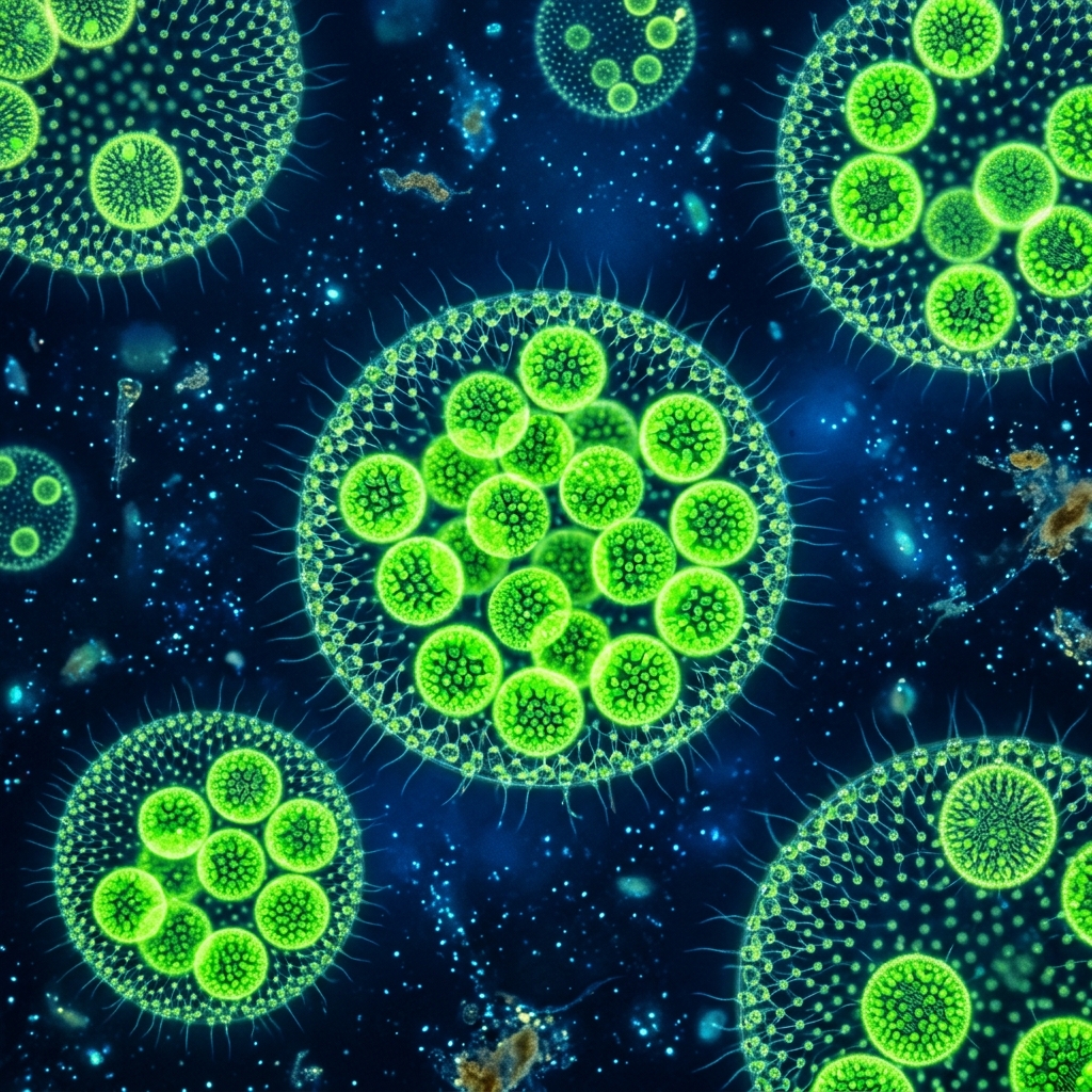
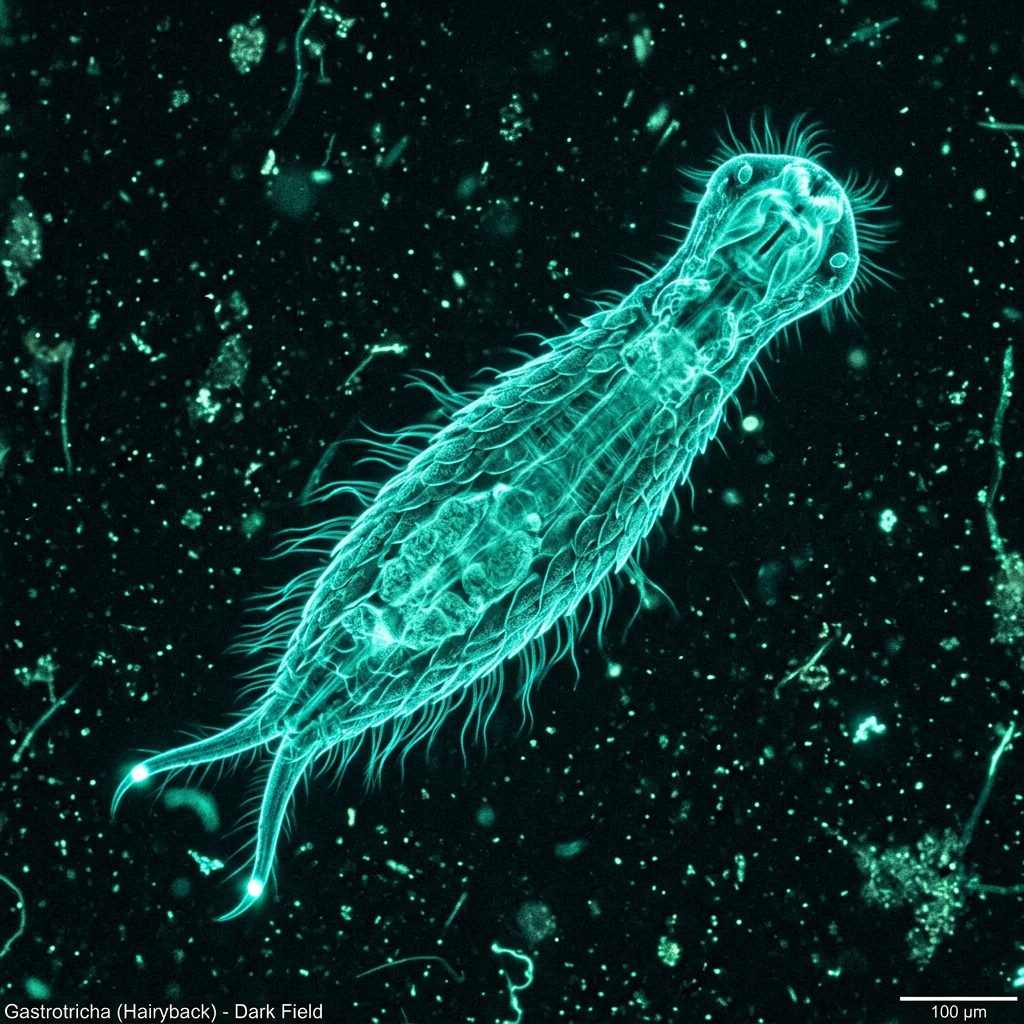
 Fig 1: A gastrotrich (*Chaetonotus*) characterized by fine dorsal spines and a furca (forked tail with adhesive tubes) (*Conceptual image).
[!NOTE] *All microorganism images used in this article are conceptual 3D CG renders.
🎯 Quick Summary & FAQ (Key Takeaways)
Before diving into the detailed guide, here are quick answers to the most common questions.
Q. Where can gastrotrichs (Chaetonotus) be found?
A. They inhabit the brown fluffy debris (detritus) at the bottom of vegetated ponds and marshes, as well as the bio-slime on aquarium glass.
If you collect pond water along with some aquatic plants and let it settle, you have a high chance of observing them scurrying along the bottom.
Q. How do they move?
A. They use the dense cilia covering their ventral surface to glide smoothly and rapidly in straight lines or wavy paths.
Unlike Paramecium, which swims in a spiraling motion, gastrotrichs always “glide” along the surface of glass or debris.
Q. What is the main challenge when observing them under a microscope?
A. They move extremely fast, so they will quickly swim out of your field of view.
You can dramatically slow them down by slightly squeezing them under the cover glass or using the “cotton fiber hack,” which traps them in a maze of cotton fibers on the slide.
🔬 1. Biology and Bowling-Pin Structure of Gastrotrichs
When looking at debris from a pond bottom under a microscope, you might spot a translucent, bowling pin- or gourd-shaped organism gliding past at incredible speeds. This is a gastrotrich, specifically from the genus Chaetonotus.
At first glance, it may look like a single-celled ciliate like Paramecium, but it is actually a multicellular micro-animal complete with a mouth, pharynx, gut, excretory organs, muscles, and a nervous system.
Spines and Adhesive Tubes: A Micro-Ninja’s Toolkit
The body of a gastrotrich is composed of several distinct regions, each serving a vital role for its survival:
- Dorsal Spines and Scales:
Its back is densely covered in countless fine spines and tile-like scales. This acts as armor against predators and serves as tactile sensors to detect environmental cues. - Cephalic Sensory Cilia:
A few long sensory hairs protrude from the sides of its head, which it extends like antennae to detect obstacles and food. - Ventral Cilia (Gastrotricha):
As the name “Gastrotricha” (stomach-hair) implies, its belly is lined with a carpet of short cilia. By beating these cilia, they glide smoothly across surfaces. - Furca (Forked Tail with Adhesive Tubes):
The tip of the tail splits like a two-pronged fork. It contains adhesive glands that secrete a sticky fluid, allowing the gastrotrich to anchor itself firmly to surfaces even in water currents.
2. Comparison with Similary Behaved Microorganisms
Here is a comparison of gastrotrichs with other elongated, surface-crawling microorganisms often encountered under the microscope.
| Feature | Gastrotrich (This species) | Nematode (Roundworm) | Paramecium (Ciliate) |
|---|---|---|---|
| Body Structure | Multicellular (head, trunk, forked tail) | Multicellular (long cylindrical, unsegmented) | Single-celled (slipper-shaped, complex organelles) |
| Body Armor | Present (dorsal spines and scales) | Absent (covered by a cuticle layer) | Absent (cilia only) |
| Movement Style | Glides smoothly using ventral cilia | Wiggles body side-to-side vigorously | Swims in a spiral path using all-over cilia |
| Tail Attachment | Yes (anchors via furcal adhesive glands) | Generally no (except some species) | No |
3. Collection Methods: Finding Them in Aquatic Plants and Mud
Because gastrotrichs are not planktonic, you won’t find many near the surface. They rely on organic debris and the surfaces of aquatic plants.
Steps from Collection to Slide Preparation
- Targeting Habitats:
Visit a local pond, marsh, or slow-moving ditch. Collect aquatic plants (like hornwort or elodea) along with the brown bottom debris (detritus) in a plastic bottle. - Aquariums as a Source:
They are also highly likely to live in the substrate of aquariums or the brown grime built up on filter sponges at home. - Sorting in a Petri Dish:
Pour your sample water thinly into a petri dish. Break up clumped debris slightly with tweezers. Under a bright light, wait a few minutes. You will likely spot tiny, ~0.2 mm gastrotrichs darting out from the debris and gliding around actively. - Slide Prep (The Cotton Fiber Hack):
Since gastrotrichs are extremely quick, they will quickly zip out of view under a standard cover slip. To prevent this, disperse a few fine strands of cotton fiber on your slide, add a drop of water containing the gastrotrichs, and place the cover glass on top. The cotton fibers create a physical maze. Trapped in these pockets, the gastrotrichs slow down, letting you observe them closely.
4. Microscope Highlights: The Pharyngeal Pump and Spiny Armor
Once the slide is ready, scan at 100x magnification to find a specimen. After spotting one trapped in the cotton fibers, switch to 400x to observe the fine details.
[!IMPORTANT] Three Key Observations & Photo Opportunities
- Anchoring & Head Swinging:
Gastrotrichs occasionally glue their forked tail to the slide, using it as a pivot point to swing their body like a compass while vigorously shaking their head left and right to search for bacteria. This is a perfect chance to lock focus and observe them.- The Powerful Pharyngeal Pump:
Right behind the head lies a robust, pear-shaped muscular pharynx. They contract this pharynx like a pump to suck in water and food, channeling it to the gut. Watching this rhythmic pumping action makes it clear that they are indeed complex “animals.”- Spines in 3D under Narrow Aperture:
Slightly closing the microscope’s condenser aperture (or using oblique illumination) makes the translucent dorsal spines and scales stand out. You can capture beautiful, 3D-like images of these creatures resembling tiny hedgehogs or armadillos.
5. Tips for Keeping and Culturing Them at Home
Here are tips to keep gastrotrichs alive in a petri dish for several weeks to a month for ongoing observation:
- Dechlorinated Water:
Chlorine is lethal. Use aged tap water (let sit for 24 hours) or commercial bottled soft mineral water. - Food Supply (Maintain Detritus):
Gastrotrichs feed on bacteria growing on detritus (organic debris). Add a tiny amount of natural pond mud or aquarium grime to the dish. - Micro-feeding with Dry Yeast:
Once every 1 to 2 weeks, dissolve a minute trace of dry yeast powder (just a speck on the tip of a toothpick—enough to avoid clouding the water) to breed bacteria. Note: Overfeeding will spoil the water and crash the culture.
With their amusing bowling-pin shapes and high-speed antics in the mud, gastrotrichs showcase a level of behavior and complexity that sets them apart from single-celled amoebas or paramecia. Give these multicellular wonders a closer look under your microscope!
🔗 Related Articles (Topic Cluster)
- 👉 Nematode Culturing & Observation: Linear Micro-Animals in Soil and Water
- 👉 Culturing & Observing Paramecium: Easy Breeding and Microscopy Tips
- 👉 Culturing & Observing Diatoms: Water Quality and Light Hacks for Glass-Shelled Micro-Gems
- 👉 The Smartphone Microscopy Guide: Three Optical Alignment Hacks to Prevent Vignetting