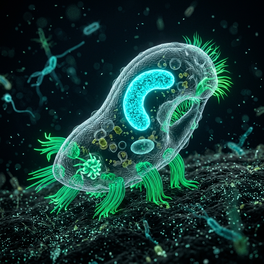
 Figure 1: Euplotes walking along the bottom of a Petri dish using its cirri like legs (*Image is for illustrative purposes only).
[!NOTE] *All microorganism images used in this article are 3D CG conceptual images.
🎯 Quick Summary & FAQ (Conclusion First)
Before diving into the detailed explanation, here are answers to the most common questions about Euplotes.
Q. What kind of organism is Euplotes? A. It is a single-celled ciliate with bundles of cilia called “cirri,” which it uses to “walk” along surfaces like slide glasses or aquatic plants. Instead of just swimming around in the water like Paramecium, they show a unique bipedal/multipedal walking action by planting their feet on objects.
Q. Where can I collect them? A. They are very commonly found in mud at the bottom of ponds and streams, around decaying leaves, or in the brown detritus (organic waste) accumulated in home aquarium filters. Areas rich in organic matter where bacteria thrive are prime hunting spots.
🔬 1. The Single-Celled “Walker”: The Amazing Anatomy of Euplotes
When observed under a microscope, Euplotes is a highly evolved single-celled organism that looks almost like an insect or a multipedal creature.
The Incredible Walking Organs: “Cirri”
The most prominent feature of Euplotes is the row of sturdy “legs” on the ventral (bottom) side of its cell. These are not single cilia, but rather bundles of dozens of cilia tightly fused together to form structures called “cirri.”
- True “Walking”: By moving these cirri in sequence, Euplotes can scramble quickly across slide glasses or cover slips, much like a spider.
- Precise Control via Microtubules: Despite being single-celled, it coordinates and controls each cirrus using an intricate internal microtubule network (cytoskeleton). This enables highly sophisticated actions, such as backing up when hitting an obstacle or changing direction, rather than just moving forward.
Large Cytostome and C-shaped Macronucleus
When observing Euplotes from directly above (dorsal side), you can see a large, funnel-shaped cytostome (cell mouth/peristome) opening on the upper left of the cell. This area is lined with bands of cilia that generate strong water currents to suck in surrounding bacteria and microalgae.
Inside the cell, there is also a huge, C-shaped or “3”-shaped macronucleus that controls the day-to-day life-support activities of the cell.
2. How to Find and Collect Euplotes
Euplotes is commonly found in freshwater environments (ponds, rice paddies, streams) in Japan and inside home aquariums.
Collection Spots and Discovery Probability
| Collection Spot | Discovery Probability | Collection Tips |
|---|---|---|
| Aquarium Filter | Extremely High (80%) | Often mixed in large numbers with other microorganisms in the brown, cloudy water and detritus squeezed from filter floss. |
| Pond Bottom Leaves or Sludge | High (75%) | Use a dropper to suck up decaying fallen leaves from the water bottom along with the surrounding mud. |
| Algae on Aquarium/Pond Glass | Moderate (60%) | Scrape off algae or fuzzy biofilm from the walls using a scraper and collect it along with the water. |
[!TIP] “Detritus” is the Key Word Because Euplotes walks along the surface of mud and organic debris (detritus) to feed on bacteria, targeting areas where some mud or debris has accumulated rather than crystal-clear water is the biggest key to a successful collection.
3. Step-by-Step Euplotes Culturing Recipe
Since Euplotes feeds primarily on bacteria, you can easily culture and multiply them long-term at home by keeping a moderate population of food bacteria.
What You Need
- Culture water containing starter Euplotes: Water collected from the wild or squeezed from an aquarium filter.
- Culture Vessel: A shallow plastic container or glass Petri dish.
- Culture Water: Dechlorinated tap water (left out overnight) or soft mineral water.
- Food (Nutrient source): 1 boiled rice grain or a tiny amount of dry yeast (less than half a pinch, just a tiny speck).
Culturing Steps
- Set Up the Vessel: Pour culture water into the Petri dish or container to a depth of about 5mm to 1cm. A flat, shallow container with a large surface area is ideal to allow oxygen to dissolve easily.
- Introduce the Starter: Use a dropper to draw water where Euplotes has been confirmed under the microscope, and add it to the container.
- Feeding: Drop a single boiled rice grain into one edge. Alternatively, dissolve a tiny speck of dry yeast in water and add just a few drops, enough to make the container water very slightly cloudy.
- Storage: Leave it in a cool place (20°C to 25°C) out of direct sunlight.
[!WARNING] Beware of Crashes from Overfeeding! If you add too much yeast or too many rice grains, bacteria will multiply explosively, depleting the dissolved oxygen and killing all the Euplotes. If the water starts to smell sour, it indicates overfeeding (anoxic state). Immediately transfer a portion of the culture to fresh water.
4. Microscope Observation and Walking Action Videography Hacks
Because Euplotes always crawls along surfaces, it is much easier to keep in focus than organisms like Paramecium that swim wildly in three dimensions. This makes it an excellent subject for beginners starting with microscope photography.
Hacks for Beautiful Observation and Photography
- Focus on the Bottom Surface: Once you prepare the slide, try focusing on the underside of the cover slip or the surface of the slide glass. You can observe Euplotes walking quietly but busily.
- Use Oblique Lighting (Angled Light): By slightly shifting the biological microscope’s condenser (diaphragm) to direct light from an angle, or partially blocking the light from below with your hand, you can create a semi-three-dimensional “oblique illumination.” This makes each individual transparent cirrus (leg) and the C-shaped macronucleus stand out sharply.
- Record Movement in Video: While still images are attractive, their true appeal lies in their walking motion. Using a smartphone microscope adapter to shoot slow-motion video allows you to clearly see how the cirri move dextrously, almost like jointed legs, resulting in highly engaging footage.
A “microscopic walking creature” that defies the conventional image of single-celled organisms. Search for them in nearby waters or aquariums, and check out their humorous walking action under the microscope!
🔗 Related Articles
- 👉 The Micro World: A Guide to Collecting and Observing Stylonychia at Home - An article on observing Stylonychia, which also belongs to the hypotrich ciliates with “cirri.”
- 👉 The Mystery of the Spring: A Step-by-Step Guide to Collecting and Culturing Vorticella at Home
- 👉 How to Culture Paramecium Easily: A Foolproof Recipe Using Rice Bran and Plastic Bottles
- 👉 Beginner’s Guide to Choosing a Home Microscope: Comparison of Recommended Models Supporting Smartphone Photography
- 👉 Smartphone Microscope Photography Guide: 3 Optical Axis Adjustment Hacks to Prevent Vignetting and Get High-Quality Images