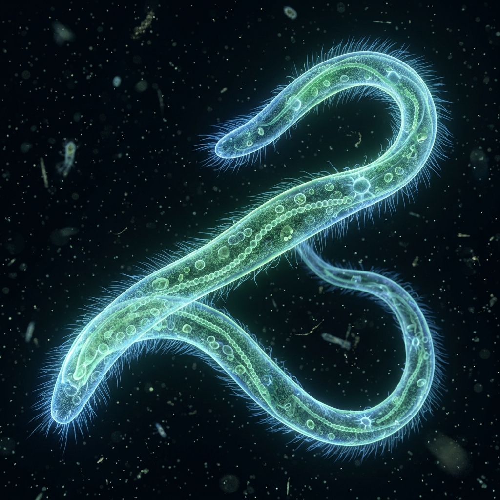
 Figure 1: Spirostomum swimming slowly with its extremely elongated body (*Image is a conceptual CG render)
[!NOTE] *All microorganism images used in this article are conceptual 3D CG renders.
🎯 Quick Summary & FAQ (Conclusion First)
Before diving into the detailed guide, let’s quickly answer some of the most common questions about Spirostomum.
Q. What kind of organism is Spirostomum? A. It is a giant single-celled organism measuring 1 to 3 mm in length, visible to the naked eye as moving white threads. Related to Paramecium (ciliates), it is characterized by its taut, slender cell body. When exposed to mechanical vibration or sudden changes in light, it contracts its body to 1/4 or 1/5 of its original length within milliseconds—one of the fastest movements in the biological kingdom.
Q. Is it difficult to culture (breed) them at home? A. No, it is quite simple. You can breed them using the same bacterial method as Paramecium, using boiled rice or Ebios tablets. However, they are more sensitive to sudden water quality degradation (lack of oxygen or excessive rot) than Paramecium. The secret to long-term culturing is to keep the water shallow to maximize the surface area exposed to air and to feed them in moderation.
🔬 1. Ecology of Spirostomum: World-Class Ultra-Rapid Contraction
Spirostomum (such as Spirostomum ambiguum, commonly found in freshwater) possesses remarkable size and motility, pushing the boundaries of what a single cell can do.
The Mystery of the Myoneme: Contracting in 0.005 Seconds
The defining feature of Spirostomum is its ultrarapid contraction. Upon stimulation, it can shrink to 25% of its original length in just 5 to 10 milliseconds (less than 1/100 of a second). This speed far exceeds the contraction velocity of animal muscle tissues. This action is not driven by actin and myosin as in animal muscles. Instead, it relies on a network of specialized contractile proteins called myonemes (mainly calcium-binding proteins like spasmin) located just beneath the cell membrane. These proteins condense instantly in response to changes in calcium ion concentration. The contracted cell then takes several seconds to dozens of seconds to slowly stretch back to its original slender shape.
Comparison of Giant Single-Celled Organisms
| Feature | Spirostomum | Stentor | Paramecium |
|---|---|---|---|
| Average Body Length | 1.0mm - 3.0mm | 0.5mm - 2.0mm | 0.15mm - 0.25mm |
| Shape | Extremely slender, cylindrical | Trumpet or horn-shaped | Slipper-shaped |
| Primary Lifestyle | Crawls on debris, swims slowly | Sessile on aquatic plants (can swim) | Constantly swims actively |
| Key Behavior | Ultrarapid spring-like contraction | Contracts from trumpet shape on contact | Backs away upon hit (avoidance reaction) |
| Visibility to Naked Eye | Extremely easy (white thread-like) | Relatively easy (green or blue specks) | Barely visible (tiny white dots) |
🧪 2. Collecting and Culturing Spirostomum at Home
Follow these step-by-step instructions to collect Spirostomum from nature and maintain a stable culture at home.
📦 Tools Needed
- Sampled water containing mud and leaves from a pond or rice paddy (best where organic matter has accumulated at the bottom)
- 1-2 grains of boiled rice or 1/4 Ebios tablet (as food for bacteria)
- Dechlorinated tap water (left out overnight) or commercial soft mineral water
- Shallow, wide container (glass petri dishes or shallow plastic food containers are ideal)
- Pipette (with a wide opening to avoid crushing the organisms)
How to Collect Spirostomum from Nature
Instead of sweeping the water column with a plankton net, you will get the best results by targeting the bottom mud and decaying leaves.
- Locate a spot: Find a stagnant pond, marsh, or the edge of a rice field where fallen leaves accumulate and the water is slightly turbid.
- Sampling: Scoop up bottom sediment, decaying leaves, and water grass along with the bottom water into a plastic bottle.
- Settling: Let the bottle sit undisturbed at home. Within a few hours to a day, Spirostomum will emerge from the mud in search of oxygen, appearing as thin, white thread-like shapes (1-3 mm) crawling on the bottom or swimming slowly near the walls.
🌿 Stable Culturing Process for Spirostomum
Since Spirostomum feeds primarily on bacteria, the basic culturing principles are identical to Paramecium. The most critical factor is using a shallow, wide container. Because of their large size, sealing them in deep bottles can lead to oxygen deprivation and toxic gas buildup, causing the culture to collapse overnight.
graph TD
    A[Prepare a shallow container] --> B[Fill with dechlorinated water to a depth of 1.5-2 cm]
    B --> C[Add 1 grain of boiled rice or 1/8 Ebios tablet]
    C --> D[Introduce Spirostomum seed using a wide pipette]
    D --> E[Place in a cool, dark room at 18-24°C]
    E --> F{Check after 1 week}
    F -->|Abundant population| G[Subculture into new containers every 2 weeks]
    F -->|Water becomes too turbid| H[Perform 50% water change to introduce fresh air]
Step-by-Step Setup
- Prepare the container: Fill a shallow glass dish or plastic container with dechlorinated water to a depth of 1.5 to 2 cm.
- Add food: Drop in one grain of boiled rice or a tiny fragment of an Ebios tablet (about 1/8 of a tablet).
- Inoculation: Use a wide-tip pipette to draw Spirostomum from your collection bottle and transfer them to the new container.
- Maintenance: Keep the container lid slightly open to ensure air exchange. Store in a cool place (18°C–24°C) out of direct sunlight.
Culturing Tips
- As bacteria multiply around the rice or Ebios tablet, Spirostomum will cluster around the bacterial colonies, appearing as white, cloudy mats.
- Water Quality: If the water becomes extremely turbid or smells foul, you are overfeeding. Use a pipette to transfer the healthy Spirostomum from the bottom to a new container with fresh water. Under normal conditions, they can be maintained by subculturing once every two weeks.
🔬 3. Microscopic Hacks for Observing Ultra-Rapid Contraction
Due to their large size, Spirostomum can be viewed in stunning detail even under low-cost educational microscopes. Use the following techniques to capture their dynamic movement.
1. The “Spacer” Hack to Prevent Crushing
With a width of about 0.1 mm to 0.2 mm, Spirostomum cells are relatively thick for ciliates. Placing a cover slip directly on top of them can crush their bodies, preventing contraction or killing them.
- Hack: Before applying the cover slip, put a tiny dab of Vaseline on its four corners, or stick small pieces of adhesive tape on both sides of the slide to act as spacers.
- Result: The cells will have enough room to swim and contract without being flattened by the cover slip.
2. Triggering Contraction via Vibration
When a Spirostomum is fully extended and stationary, lightly tap the edge of the slide or the microscope stage with a pen or finger to create a vibration.
- What to watch: The instant you tap, the elongated Spirostomum will shrink into a short, rounded egg or eggplant shape. You can then watch it slowly stretch back into a thread over 10 to 30 seconds.
3. Slow-Motion Videography with a Smartphone
Because Spirostomum contracts in less than 1/100 of a second, standard video recording (30 fps or 60 fps) will only capture one or two frames of the actual contraction, making it look like they teleported.
- Hack: Mount your smartphone camera to the microscope eyepiece (using a smartphone microscope adapter makes this easy) and record in “Super Slow Motion” mode (240 fps to 960 fps).
- Result: When played back, you can see the cell twist and compress in slow motion, propagating from one end to the other—revealing the physical mechanics of this cellular contraction.