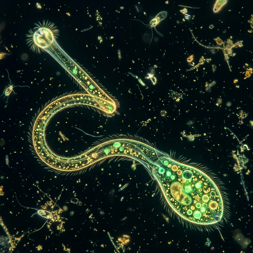
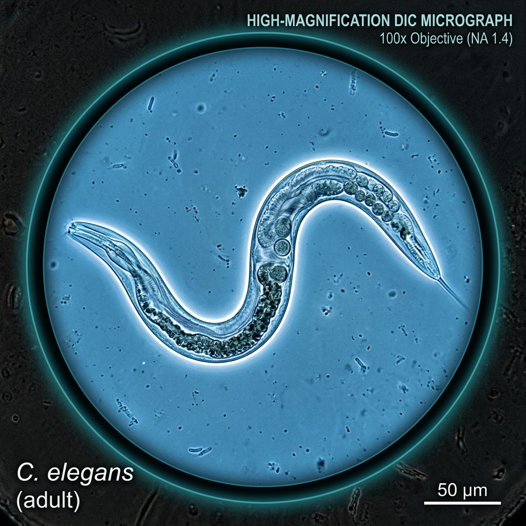
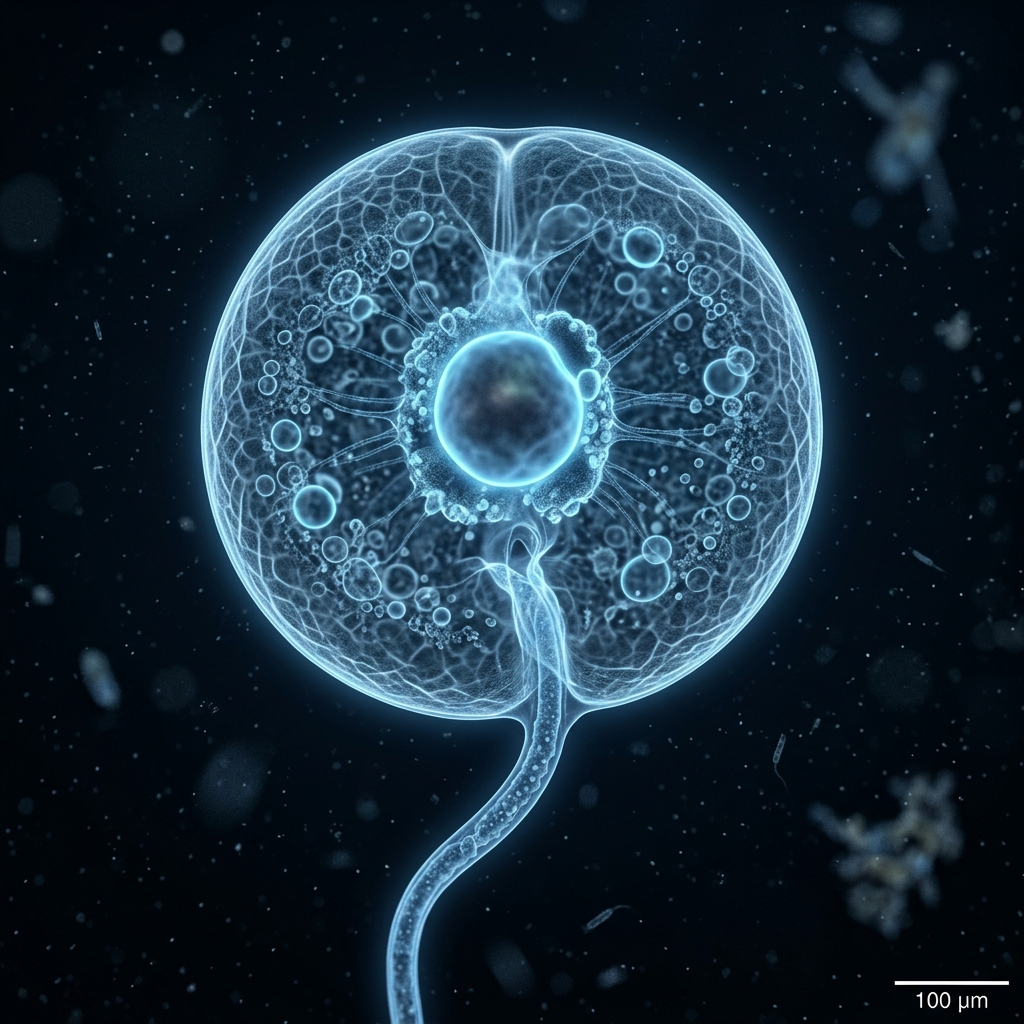
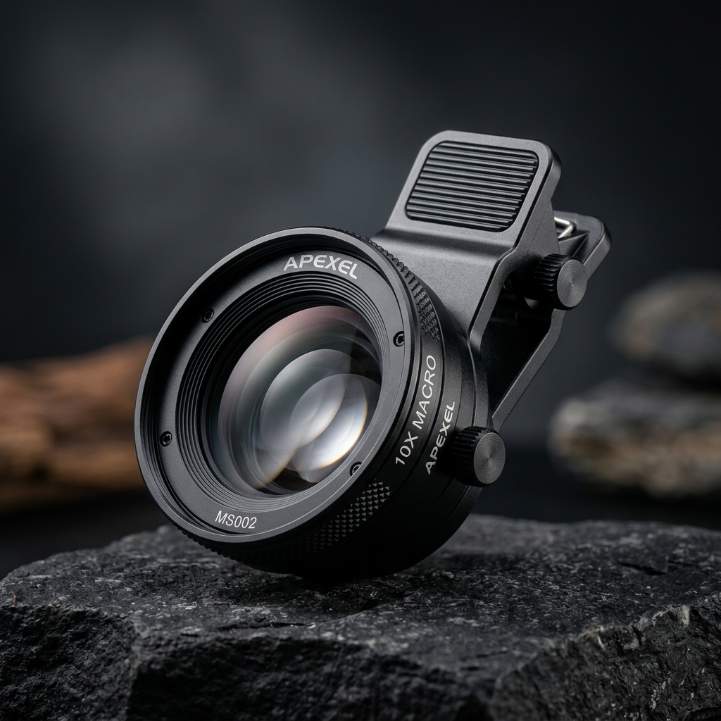
 Figure 1: Layout diagram of the microscope imaging system (*Image is for illustrative purposes only)
[!NOTE] *All images used in this article are illustrative images.
In microscopy, the higher the magnification, the thinner the “depth of field” (the slice of the specimen that remains in sharp focus)—reducing it down to the sub-micron level. “Focus stacking” (or depth composition) is a software-based technique to solve this physical limitation.
Capture and Stacking Procedure
- Clamp and lock your smartphone completely still. Start with the microscope focus dial set at the closest edge of the specimen.
- Slowly turn the focus dial to shift the focal plane slightly deeper, taking one picture at each step (we recommend 10 to 20 shots).
- Import your sequence of photos into a free image stacking program (such as CombineZP or Fiji/ImageJ).
- Run the focus stacking command. The software will automatically detect and extract only the sharpest parts of each frame and merge them together, rendering a fully focused, three-dimensional view of the entire microorganism.