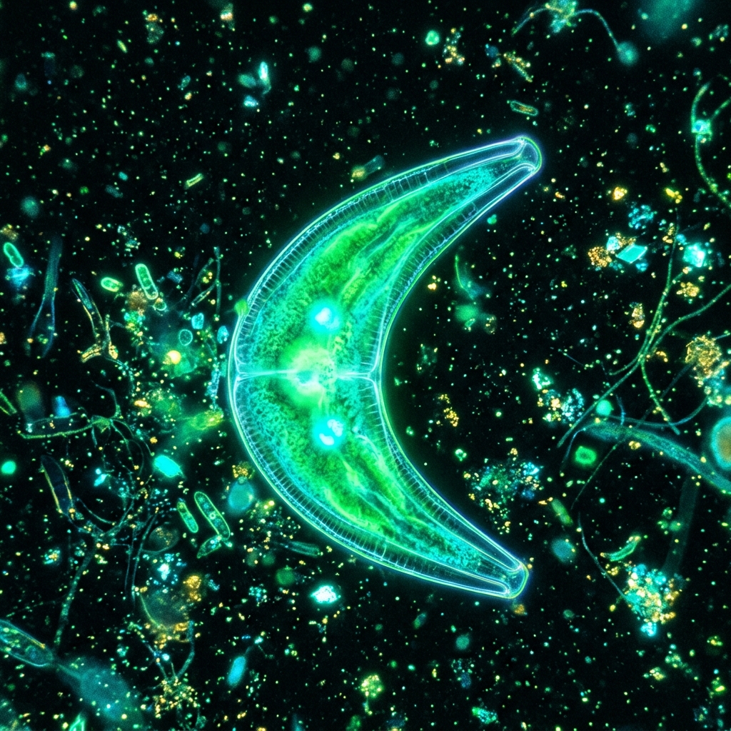
 Fig 1: A Closterium cell exhibiting a graceful mirror-symmetry, with its central nucleus and large flanking chloroplasts (*Conceptual image).
[!NOTE] *All microorganism images used in this article are conceptual 3D CG renders.
🎯 Quick Summary & FAQ (Key Takeaways)
Before diving into the detailed guide, here are quick answers to the most common questions.
Q. Where can Closterium be collected?
A. They commonly inhabit sunny rice paddies (especially after planting), old ponds, and marshes, particularly in bottom mud and around aquatic plants.
If you collect pond water along with bottom sediment in a transparent bottle and place it near a window, they will gather toward the light due to positive phototaxis, allowing you to isolate them with a pipette.
Q. Why do the tips of the cell appear to shake under the microscope?
A. Tiny calcium sulfate crystals inside vesicles at both tips (terminal vacuoles) vibrate randomly due to Brownian motion.
This is not active movement by the organism, but a physical phenomenon where water molecules collide with the tiny crystals (Brownian motion). It is a highlight of observing Closterium. At 400x magnification or higher, you can clearly see these crystals dancing inside their terminal chambers.
🔬 1. Cellular Structure and Physiology of Closterium
Closterium is a single-celled green alga (conjugating green alga). Despite being a single cell, its geometry is exceptionally beautiful, making it a classic subject for microscopic observation.
Perfect Mirror-Symmetry
The cell is divided by a central constriction called the isthmus, splitting the cell into two matching halves (semicells) that are mirror images of each other.
- Isthmus (Center): The location of the nucleus. During asexual division, the cell splits here, and each half regenerates the missing semicell.
- Chloroplasts: These occupy most of the semicells, displaying a star- or ribbon-like green structure. They capture light to photosynthesize starch. Within the chloroplasts, starch-storing bodies called pyrenoids can be seen arranged in a neat row.
The Crystal Dance: Brownian Motion in Terminal Vacuoles
At the pointed tips of the cell are round vacuolar compartments called terminal vacuoles. Looking closely, you will see tiny translucent crystals shimmering and vibrating erratically.
These particles are calcium sulfate (gypsum) crystals, formed as a byproduct of cell metabolism or waste excretion. Elevating the magnification to 400x–600x and focusing on the tips reveals a real-time display of Brownian motion as water molecules bombard the crystals. It is a stunning intersection of biology and physics.
🧪 2. Foolproof Recipe for Closterium Culture Medium
Since Closterium is a photosynthetic autotroph, it does not require organic food (like bacteria or rice grains). Adding organic matter will cause bacterial and fungal blooms, leading to water spoilage and culture crashes.
Like hydroponic plants, they require very dilute inorganic nutrients (minerals) and adequate light to thrive on your desk.
📦 What You Need
- Closterium starter culture (collected from rice paddies or purchased online)
- Aged, dechlorinated tap water (let tap water sit under light for 24 hours to remove chlorine)
- Liquid garden fertilizer (e.g., Hyponex liquid)
- 200 mL–500 mL transparent container (glass petri dish, clean jar, or clear plastic bottle)
- LED grow light or a bright windowsill out of direct sun
🌾 Preparing DIY Medium (1:10,000 Fertilizer Dilution)
While academic labs use sterile “C-medium,” hobbyists can substitute it with a highly diluted liquid fertilizer solution, a recipe also used for culturing Volvox.
Step 1: Prepare the Water
Pour 1 liter (1,000 mL) of dechlorinated water into a clean container.
Step 2: Add Fertilizer (1:10,000 to 1:20,000 dilution)
Use a dropper to add exactly 2 drops (approx. 0.1 mL) of liquid fertilizer.
[!WARNING] Avoid the temptation to add more fertilizer! Excess nutrients will cause green water blooms or filamentous algae (like Spirogyra) to overgrow the walls, outcompeting and starving the Closterium.
Step 3: Inoculate and Set Up
Add 10%–20% volume of Closterium starter water to the prepared medium and stir gently. Place the lid loosely so carbon dioxide can enter. Position the vessel 15–20 cm away from the LED grow light and illuminate for 10–12 hours a day.
3. Troubleshooting & Health Maintenance Matrix
By monitoring your culture container, you can keep a colony of Closterium thriving for months.
| Sign / Observation | Potential Cause | Action Plan / Fix |
|---|---|---|
| Cells turn pale green or yellowish | Light deficiency or nutrient depletion (nitrogen/phosphorus) | Extend the light exposure time slightly, or add a half-to-one drop of fertilizer. |
| Brown slimy film forms on the bottom or walls | Overgrowth of diatoms or other unwanted plankton | Pipette out the clean green Closterium layer from the bottom and transfer them to a clean bottle with fresh medium. |
| Water turns cloudy white with a foul smell | Organic contamination leading to bacterial spoilage | Closterium is highly sensitive to poor water quality. Promptly isolate healthy cells and transfer them to fresh medium in a clean container. |
| Cells become overly curved, shriveled, or deformed | Osmotic stress due to evaporation concentrating salts/ions | Replenish the lost volume back to the original level with pure dechlorinated water (or distilled water) without any fertilizer. |
🔬 4. Microscope Highlights: Conjugation and Zygospores
When observing Closterium under the microscope, fine-tuning your illumination and magnification options reveals biological features beyond basic text-book drawings.
Observing Gliding Movement at the Tips
Although they seem static, Closterium cells move. They perform a gliding movement toward light by secreting thin, invisible mucilage through tiny pores at their tips. If you watch a cell under the microscope over several minutes, you may notice it slowly shifting direction or sliding across the field of view.
Capturing Conjugation (Sexual Reproduction)
When the culture begins to dry out or nutrients shift, Closterium cells initiate conjugation as a survival mechanism.
- Alignment: Two different cells align side-by-side in close proximity.
- Conjugation Tube: Both cells extend a small bulge from their central isthmus, forming a connecting tube.
- Protoplast Fusion: The cellular contents emerge from the cells and fuse into a single mass inside the tube.
- Zygospore Formation: The fused protoplast develops a thick, protective wall (often star-shaped or polygonal) to become a zygospore. This spore can withstand drying and freezing temperatures, remaining dormant for years until conditions improve.
The distinctive, geometric zygospore is a highly prized sight for microscopy enthusiasts.
🔗 Related Articles (Topic Cluster)
- 👉 How to Choose a Home Microscope: A Foolproof Selection Guide with Smartphone Photography Comparison
- 👉 The Smartphone Microscopy Guide: Three Optical Alignment Hacks to Prevent Vignetting
- 👉 Overcoming Depth of Field: Focus Stacking Hacks for 3D Microscope Photos with Free Software
- 👉 Culturing Volvox & Observing Asexual Reproduction: DIY Medium and Key Points
![[Microscopic Photography] Master Pseudo-Koehler Illumination Adjustment: The Ultimate Hack to Beautifully Record Microscopic Details](/images/tool_ring_light.png)
![[Microscopic Photography] Master Stage Micrometer Calibration: The Ultimate Hack to Beautifully Record Microscopic Details](/images/microscope_clamp.png)
![[Microscopic Photography] Master Glycerin Mounting: The Ultimate Hack to Beautifully Record Microscopic Details](/images/tool_microscope_stand.png)